 Login
LoginX-Ray (General Radiology and Fluoroscopy)

- Introduction to x-rays
- How do x-rays work?
- Preparing for an x-ray
- What happens during an x-ray?
- Risks and benefits of x-rays
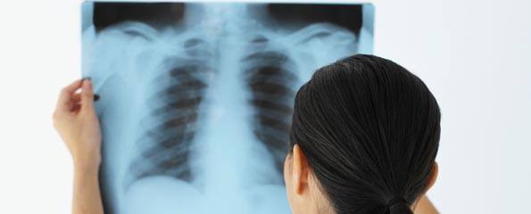
Introduction to x-rays

The most common and oldest form of radiology, the x-ray is the common key behind many radiological procedures.
A fairly simple process, an x-ray involves the x-rays being emitted by the machine as particles that pass through the body, being absorbed at different frequencies by different internal structures until they are detected by the sensitive film, which produces the image.
The x-ray produces images that resemble the shadow of internal structures such as bone and tissue.
How do x-rays work?
An x-ray is a procedure fairly easy to comprehend. Discovered in 1895 by German physicist Wilhelm Roentgen, by accident, Roentgen experimented with electron beams in a gas discharge tube and discovered the benefits of seeing the shadow of bones and tissue through the body.
Basically the process of producing an x-ray involves a source that produces x-rays and radiating them through a body or object, where they are absorbed at different rates until they are detected by a sensitive film or cassette.
The x-ray is a form of electromagnetic energy, similar to light rays, but of a higher energy level and different wavelength which allows them to pass through the body and create internal images. The bodily structures absorb these x-rays at different rates, for example, bones are very dense and absorb a larger amount of x-rays, whereas the surrounding tissue is less dense and absorb less x-rays. The resulting images are the different levels of absorption of these x-rays.
In fluoroscopy, the x-rays pass through the body onto a fluorescent screen, creating a moving X-ray image. Doctors may use fluoroscopy to trace the passage of contrast media through the body. Doctors can also record the moving X-ray images on film or video. Today the ability to switch between conventional and fluoroscopic techniques is available, with the enhancement of digital imaging.
Preparing for an x-ray
Preparation for an x-ray is very similar to other radiological procedures. There are a few points that are useful to know before you attend the procedure.
- It is recommended that you wear loose fitting clothes, but commonly you will be asked to undress partially or to wear a hospital gown.
- All jewellery, belts, hats or anything with metal components will have to be removed, as they distort the picture (this includes bras, hair pins/clips, and clothes with metal components like zippers); it is recommended that you leave these items at home for safe keeping.
- Arrive at least ten minutes early for the procedure to allow for filling out of forms etc.
- Inform your doctor before booking the procedure or radiologist, technician or nurse before the procedure if you think you may be pregnant or you are pregnant as this procedure does involve radiation that could damage a developing foetus.
What happens during an x-ray?
Once you have filled out the necessary forms, the radiologists will call you through where you will either be asked to put on a hospital gown or you will be led into the procedure room. During this time, the radiologist will ask for you to remove any objects or articles that may distort the picture.
Some x-rays require a contrast agent being administered to allow for a clearer picture of certain tissues and internal structures.
The radiologist or technician will then advise you of the correct position which you will stay in during the procedure to allow for the particular part of the body that is of interest to imaged clearly. The radiologist or technician then leaves the room, or steps behind a screen so they are not exposed to the radiation produced.
You will be asked to remain very still and during the imaging you may be asked to hold your breath to allow for clarity of the picture. The procedure usually takes up to thirty minutes.
After the x-rays are completed you will be asked to redress and wait until the images are processed and reviewed by the radiologist. You may then be required to re-take some x-rays, or you will be released. The x-rays generally are either sent to your doctor, or they may require you to collect them from the radiology clinic. An appointment can then be made with your doctor to review the results.
Risks and benefits of x-rays
As x-rays are a procedure that uses a radioactive component to produce the images, there is risk involved. However this risk is very minimal due to the efforts of radiologists to reduce the amount of radioactivity being produced. Compared to the early days of x-ray technology, the amount being used is greatly reduced. Often the use of a lead apron or belt protects the other areas of the body from exposure, as lead is a dense metal that absorbs 100% of the radiation produced.
Technology today means that advanced x-ray techniques reduce the area being exposed and subsequent scatter radiation and the amount being used to produce the images. The diagnostic benefits that are associated with ability of x-ray outweigh the very minimal possibility of any issues arising from an x-ray procedure.

Dates
Tags
Created by:


