 Login
LoginEye examinations (visual field testing)

- Introduction to eye examinations
- Who should have their eyes examined?
- How are eye examinations conducted?
- What eye disorders are detected through eye examination?
- How effective are eye examinations in preventing eye disorders?
Introduction to eye examinations
 Eye examination, also known as visual field testing, is the process of having the eyes examined for visual field defects or for monitoring changes in vision. Having the eyes examined regularly is the best way to detect vision changes and ensure that the conditions underlying changes in vision are diagnosed early.
Early diagnosis enables early intervention and treatment of emerging conditions, which prevents worsening of symptoms (e.g. vision loss). As many eye diseases remain asymptomatic until they have progressed significantly, eye examinations are often the only means of detecting eye problems before irreversible vision loss has occurred.
Who should have their eyes examined?
Everyone is exposed to some of the factors which make eye disease more likely (e.g. sunlight) and thus everybody should have their eyes comprehensively examined, periodically, by an ophthalmologist. Adults with no apparent risk factors (outlined below) or visual defects should commence visual field testing once they reach 40 years of age.
The American Academy of Ophthalmology recommends eye examinations at different frequencies, depending on the individual’s age:
- Aged 40-54 years: every 2-4 years;
- Aged 55-64 years: every 1-3 years;
- Aged 65 years or more: every 1-2 years.
Minor eye examinations may be necessary to investigate specific complaints in the interim period. Individuals should also have an eye exam if they notice changes to their vision.
Individuals with a high risk of eye disease
Some individuals have a higher risk of developing visual field abnormalities than others, and these people should visit an ophthalmologist for more frequent eye examinations, as outlined below:
- Diabetes mellitus type 1: The first comprehensive eye examination should be conducted five years after diabetes onset, with yearly examinations thereafter;
- Diabetes mellitus type 2: The first comprehensive visual field test should be conducted at diabetes diagnosis, with yearly examinations thereafter;
- Pregnancy: Women should have a visual field test prior to or in the first trimester of pregnancy and every three months thereafter if risk factors for eye disease are identified;
- High risk of glaucoma: Including individuals with a family history of the condition, those of African or Hispanic origin should have their eyes examined twice as frequently as other individuals in their age category (e.g. those aged over 65 years should have 6-12 monthly examinations);
- History of trauma: Individuals with history of trauma to the eye may require more frequent examination;
- Eye abnormalities: Individuals with abnormalities identified through previous examination may also need more regular testing.
Children
 Children should be examined more frequently than adults while their eyes are still growing. Many interventions can successfully correct minor vision disorders if they are implemented while the eyes are still developing (i.e. before eight years of age). For example amblyopia, a condition in which one eye is dominant in vision and the other is lazy, can be treated with simple measures such as patching the dominant eye, as long as the eyes are still developing. However, once a child reaches eight years of age the condition is unlikely to be resolved, even following intensive treatment. Individuals with untreated amblyopia are at increased risk for vision loss later in life, and so childhood interventions also have an impact on the vision of the adult.
There are also significant associations between vision problems and learning difficulties in children, and thus vision problems may irreversibly affect a child’s intellectual development. Common childhood eye disorders which are likely to go undetected in the absence of visual field testing include strabismus (issues with processing of binocular images) and refractive errors (i.e. short or long sightedness or both).
Refractive errors are particularly common, and uncorrected refractive errors cause visual impairment or blindness in an estimated 153 million people worldwide. In Australia, a study based in Victoria reported that refractive error is responsible for some 8% of blindness. In addition some 23% of people who are legally blind have lost vision due to uncorrected or under-corrected refractive error.
Recent evidence regarding refractive errors and spectacle use in Australian children suggests a much lower prevalence of uncorrected refractive error. A 2006 study of Australian children reported that 10% with refractive errors were untreated (i.e. were not wearing prescription glasses). This is a relatively low proportion of children, compared to other developed countries. For example, in the United States around 30% of children have refractive errors and an estimated 90% of children in the US who require prescription glasses are not wearing them.
In children, visual assessment should typically be performed at six months, three years and at the commencement of schooling. Visual assessment should be conducted every two years thereafter until the child reaches 16 years. In children who have known risk factors for eye disorders (e.g. family history) eye examination should be performed more regularly.
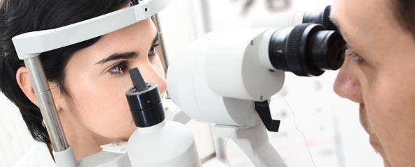
How are eye examinations conducted?
An eye examination is a process where the health professional gathers relevant background information from the person being tested, examines the eyes and implements management strategies for individuals with disease or who are likely to develop disease.
An eye examination will typically begin with the doctor asking a series of questions known as the medical history. The questioning is likely to pay particular attention to any history of eye disease, disorders and injury, as well as family history of eye disease and current vision status.
The next step is usually a comprehensive eye examination which is likely to investigate the following features of the individual’s vision:
- Visual acuity, i.e. how well and how far the patient can see. The test is usually performed with and without glasses, for patients who wear glasses;
- External examination of the eyes including the lids, lashes and lacrimal structures (tear ducts);
- Assessment of the eyes’ alignment;
- Examination of the surface of the eyes;
- Measurement of internal pressure of the eye;
- Examination of the fundus (or base) of the eye, including retina, vitreous and optic nerve.
A range of other tests (e.g. laboratory tests for systemic disease, colour vision screening) may also be conducted, depending on the findings of the standard comprehensive eye examination. These are not however routinely performed in all patients. Following the eye examination, the practitioner will generally recommend a strategy for managing the patient’s eye health in the future, which may involve scheduling a follow up visit, or an active intervention (e.g. glasses), depending on the finding of the eye exam.
In children, the types of tests conducted will depend on the age, visual and intellectual development of the child. However, tests exist which can be used even in newborn babies and toddlers.
What eye disorders are detected through eye examination?
 Eye examination is performed to detect visual abnormalities which may lead to visual loss as well as risk factors for eye disease. Appropriate interventions can then be initiated to manage those conditions and prevent vision loss. Eye examination is also used to monitor changes to the visual field in patients with deteriorating vision. Abnormalities identified through eye examination often indicate disorders of the eye but may also indicate underlying disease affecting other body systems (e.g. diabetes, AIDS). The key eye disorders which typically remain asymptomatic for extended periods but might be detected by an eye examination are outlined further below.
Glaucoma
Glaucoma refers to a range of eye diseases which cause damage to the optic nerve, or the nerve which transmits images from the eye’s retina to the brain. Damage to the optic nerve is irreversible and blindness results once the entire nerve becomes damaged. Glaucoma can be further categorised as open or closed angle. It is the leading cause of blindness globally and one of the leading causes of blindness in Australia (following cataract).
Diabetic eye disease
Diabetic eye disease refers to a group of eye conditions which arise in diabetics as a result of the affect of the disease on the small blood vessels of the eye. Diabetic proliferative retinopathy is the most common eye condition associated with diabetes. Changes to the retinal capillary system resulting from diabetes cause aneurysm in some vessels and occlusion in others.
It threatens the sight of some 60% of diabetics after 20 years of disease and, in the past, has caused blindness in some 5% of diabetics after 30 years. Blindness results in about 50% of cases of proliferative retinopathy if the condition is left untreated. Diabetic retinopathy can also lead to diabetic maculopathy, a condition which begins with macular oedema and can cause blindness if left untreated. Diabetics have a higher risk of developing cataracts earlier in life.
In Australia, an estimated 7.5% of the adult population has diabetes, and the majority of these will develop diabetic eye disease within 20 years. This has the potential to cause significant vision loss and blindness, if diabetic eye diseases are not detected and treated before irreversible ocular damage occurs.
Age related macular degeneration
Age related macular degeneration or maculopathy is an acquired eye disorder. There is no cure and it is the most prominent cause of age related blindness in Australia. It is a neurodegenerative condition, affecting the central retina (or macula) which enables high acuity vision (i.e. the clear, sharp vision necessary for reading, driving and other such tasks).
Cataracts
Cataract is an eye condition characterised by the lens of the eye becoming increasingly opaque or cloudy. It leads to impaired eyesight and in severe cases blindness. Globally, it is a major cause of vision impairment, which mainly affects elderly people.
Cataracts are prevalent in Australia; 160,000 are treated each year. The Blue Mountains Eye Study, conducted in NSW, Australia, found that the annual incidence of cataracts was 0.3% amongst individuals aged 49-54, 1.7% for those aged 55-64 years, 7.9% in the 65-74 year age group and 17.4% in individuals aged more than 75 years.
How effective are eye examinations in preventing eye disorders?
Eye examination involves testing the eye’s visual field and then implementing appropriate strategies to prevent visual field deterioration (i.e. vision loss). As damage to the eyes is typically irreversible, implementing preventative interventions early in the course of disease is essential for preventing impaired vision. Estimates suggest that up to 40% of vision loss experienced by aged home residents could be prevented if properly managed in the early stages of disease. Proper management can only be implemented if eye diseases are identified in their early stages, and thus eye examination plays a key role in the prevention of eye disorders.
Eye examinations alone do not prevent eye disorders, but the findings from an eye examination can be used to institute appropriate management strategies early in the course of an eye disease, before irreversible damage is done. For example, individuals may be identified as being at high risk of age-related macular degeneration through an eye examination and these individuals may delay the progression of their disease by taking high dose anti-oxidant supplementation and quitting smoking.
References
- Optometrists Association of Australia. Visual field testing. 2005. [cited 2009, May 15] Available from: [URL]
- American Academy of Ophthalmology Preferred Practice Patterns Committee. Preferred Practice Pattern Guidelines. Comprehensive Adult Medical Eye Evaluation. San Francisco, CA: American Academy of Ophthalmology. 2005. [cited 2009, May 15] Available from: [URL]
- Dunlop, C. Is it time to review the screening guidelines for young diabetic children? MJA 2006;184(9):476.
- American Academy of Ophthalmology Panel on Pediatric Eye and Vision Examination. Optometric Clinical Practice Guideline: Pediatric Eye and Vision Examination American Academy of Ophthalmology. 2002. [cited 2009, May 15] Available from: [URL]
- Holden, B.A. Undercorrected refractive error: the major and most easily avoidable cause of vision loss. Com Eye Health J. 2007;20(63):37-8
- Taylor, H.R. Refractive Errors: Magnitude of the need. Com Eye Health J. 2000;13(33):1-2
- Robaei, D. Kifley, A. Rose, K.A. Mitchell, P. Refractive error and patterns of spectacle use in 12 year old Australian children. Ophthalmology. 2006;113(9):1567-73.
- National Eye Institute. Glaucoma. National Institutes of Health United States, 2009. [cited 2009, May 15] Available from: [URL]
- Foran, S. Wang, J.J. Mitchell, P. Causes of incident visual impairment: the Blue Mountains Eye Study. Arch Ophthalmol. 2002;120:613-9.
- Kumar, P. Clark, M. Clinical Medicine, 5th ed, Edinburgh: Elsevier Science Ltd; 2002.
- Jackson, C.L. Hirst, L. Jong, I. Smith, N. Can Australian general practitioners effectively screen for diabetic retinopathy? A pilot study BMC Family Practice. 2002;3:4-10.
- Chiu, C. Milton, R.C. Gensler, G. Taylor, A. Association between dietary glycemic index and age-related macular degeneration in non-diabetic participants in the age-related Eye Disease Study Am J Clin Nutr. 2007; 86:180-8.
- World Health Organisation, Global disease burden from solar ultraviolet radiation. 2006, [cited 2009, April 1] Available from: [URL]
- Australian Cancer Council. Eye Protection from Ultraviolet Radiation. 2006. [cited 2009, April 1] Available from: [URL]
- Panchapakesan, J. Mitchell, P. Tumuluri, K. et al Five year incidence of cataract surgery: the Blue Mountains Eye Study. Br J Ophthamol, 2003;87:168-72.
Dates
Created by:


