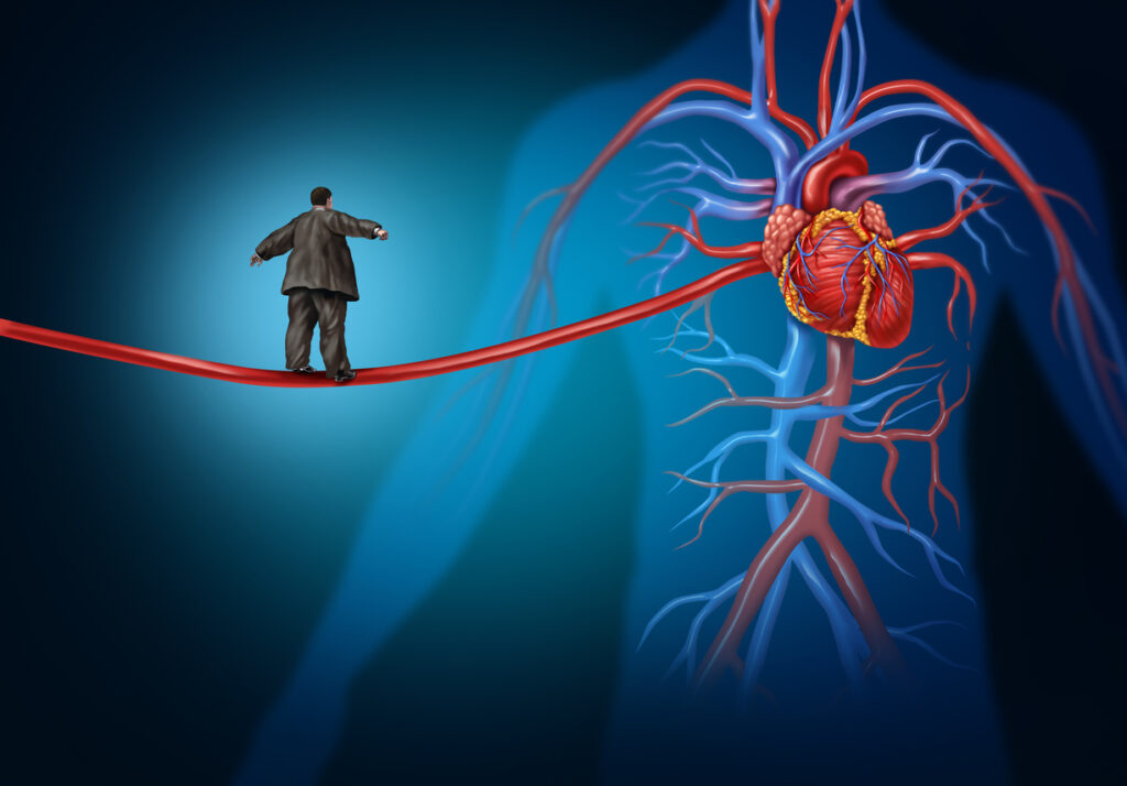
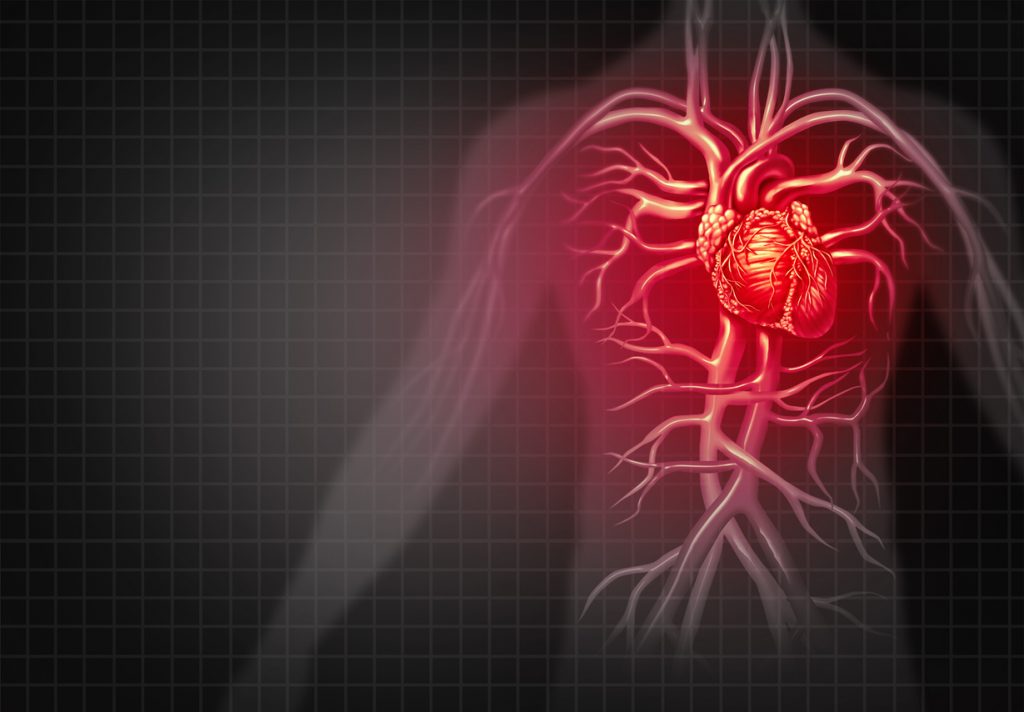
This study will examine how heart stiffness and a weak atrium affect exercise capacity and symptoms in patients with hypertrophic cardiomyopathy (HCM). The atrium is the booster pumping chamber of the heart that helps the ventricle (main pumping chamber), to fill properly. HCM is an inherited disease in which the ventricle becomes thickened and, in some patients, stiff. The stiffness makes it difficult for the ventricle to fill and empty, causing breathing difficulty, fatigue, and reduced exercise capacity. Scar formation and a weakened atrium can cause the heart to stiffen. Information gained from this study may guide doctors in prescribing medicines to reduce scarring or improve atrial function.
Official Title
The Role of Atrio-Ventricular Coupling in Exercise Tolerance in Non-Obstructive Hypertrophic Cardiomyopathy
Conditions
– Cardiomyopathy, Hypertrophic
Study Type
Observational
Study Design
Natural History
Further Details
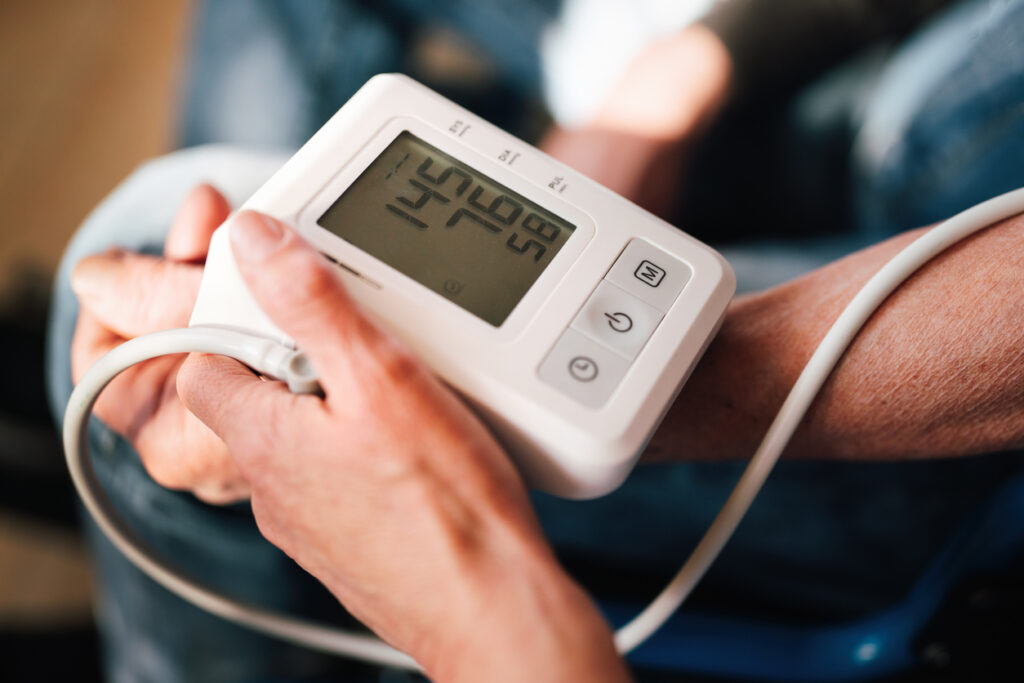
Patients 21 years of age and older with hypertrophic cardiomyopathy may be eligible for this study. Candidates will be screened with a medical history and physical examination, electrocardiogram (EKG), blood tests, Holter monitor, and echocardiogram. A Holter monitor is a device about the size of a Walkman that is connected to three wires that are attached to the chest. It is worn for 24 hours to provide continuous monitoring of heart rhythm. An echocardiogram uses a small probe that emits sound waves to produce images of the heart. The probe is moved across the chest and the reflection of the sound waves from the chambers of the heart produce images showing the heart’s thickness and function. Participants will undergo the following tests and procedures over 3 days:-Physical examination and echocardiogram.-Intravenous cannula insertion: A plastic tube is inserted into an arm vein for collecting blood samples to measure substances that the heart and circulatory system release at rest and during exercise. -Impedance cardiography: A small current of electricity is passed across the chest and electrodes similar to those used for an EKG test are placed to measure blood flow in the area of the current. -Pulmonary artery catheterization: A catheter (plastic tube) is inserted into a vein either in the arm, under the collarbone, or in the neck and advanced to the right atrium and ventricle. The catheter remains in place during the echocardiogram tilt and bicycle exercise tests (see below). -Echocardiogram tilt test: The patient lies flat on a table. After a few minutes, the table is tilted so that the patient’s head is just above his or her feet for a short while, then is positioned flat again, and then tilted so the feet are just above the head. Echocardiographic measurements and blood samples are taken at intervals to examine heart function during changes in posture. -Echocardiogram bicycle stress test: The patient exercises for as long as possible on a bicycle-like machine while lying on his or her back. Echocardiographic measurements and blood samples are taken at intervals during the test. -Treadmill stress test: The patient runs for as long as possible on a treadmill that increases in difficulty. The patient wears a facemask or mouthpiece through which small amounts of gases are added in order to measure the ability of the heart and lung to increase their effectiveness with exercise. -Digoxin loading: Only patients who demonstrate limited exercise capacity and for whom digoxin is not a risk will undergo this procedure. A medicine that makes the heart contract more strongly, digoxin is used to treat certain heart abnormalities. Patients are given doses of either digoxin or placebo (a look-alike injection with no active ingredient) at 4-hour intervals over a 24-hour period and then repeat the tilt test and the bicycle and treadmill exercise tests.Primary hypertrophic cardiomyopathy (HCM) is a genetic cardiac disease characterized by thickening (hypertrophy) of the left ventricular (LV) wall, dyspnea and/or fatigue in the setting of a normal or supra-normal LV ejection fraction. The specific mechanisms underlying heart failure-related symptomatology in non-obstructive HCM are poorly defined, but as the vast majority of HCM patients with heart failure have apparently preserved LV contractile function, their symptoms of dyspnea and fatigue are presumed due to perturbations of the relaxation/filling phase (diastole) of the cardiac cycle, which has been termed “diastolic dysfunction”. In fact, diastole is mechanistically complex and involves LV pressure decay (relaxation), chamber compliance and atrial contractile function. LV end-diastolic volume, which represents fiber stretch, governs LV contractile function and stroke volume via the Frank-Starling mechanism. End-diastolic fiber stretch is, in turn, dependent on late diastolic filling due to atrial ejection. This atrial “booster pump” is load-dependent and also responsive to inotropic effect. The interaction of atrial inotropic reserve, LV end-diastolic pressure (atrial afterload) and LV compliance (which mediates LV end-diastolic pressure and volume) may be generically considered as “atrio-ventricular coupling” which, in theory, should be at least partially responsible for modulations in exercise-induced augmentation of cardiac output related to enhancement of LV end-diastolic volume or “preload reserve”. Previous studies have suggested that limitations of preload reserve may explain exercise-associated symptoms of congestive heart failure. The potential ability of new technologies to accurately assess atrio-ventricular coupling as it relates to preload reserve present opportunities for investigation into mechanisms of heart failure operative in patients with stiff left ventricles with intact systolic function. Elucidation of these previously unapproachable mechanisms may be important in targeting therapy and the design and analysis of future interventional trials. In this pilot study, we hypothesize that exercise intolerance in HCM patients is due to limited LV preload-reserve which, in turn, is mediated by disequilibrium of atrio-ventricular coupling and, possibly, limitations in atrial inotropic reserve. We will test novel analytic tools, including measures of LV compliance and load-independent atrial systolic fuction (atrial systolic elastance), in attempts to dissect out the components of atrio-ventricular coupling which underly HCM-associated symptoms and reduced preload reserve. Further, we will assess serum and cardiac MRI markers of myocardial fibrosis to determine the effect of collagen remodeling on LV relaxation, compliance and atrial afterload. Finally, we will examine the effects of short-term cardiac glycoside (inotropic) therapy on atrial systolic elastance, preload reserve and exercise tolerance. The results of this investigation will be implemented in the design of subsequent interventional protocols targeted towards mechanisms of the stiff heart syndrome.
Study Start
Eligibility & Criteria
Genders Eligible for Study: Both Criteria INCLUSION CRITERIA – HCM Patients:HCM defined as maximal LV wall thickness by echocardiography greater than 13mm in the absence of other causes of LVH or greater than 15mm asymmetrical LV wall thickness if there is a history of mild hypertension (defined as systolic less than 160mmHg and diastolic less than 100mHg) controlled for greater than 6 monthsNon-obstructive HCMAge greater than or equal to 21 years.Patients with LV obstruction treated by LV myotomy and myectomy or percutaneous septal alcohol ablation that meet inclusion criteria are eligible for this study.EXCLUSION CRITERIA – HCM Patients:LV outflow obstruction noted during Doppler echocardiography at rest or with Valsalva maneuver defined as instantaneous peak gradient greater than 30 mmHgHemodynamically significant valvular disorders, history of significant coronary obstruction (greater than 50% in any single artery), angina symptoms, myocardial ischemia on an imaging stress test or evidence of prior myocardial infarctionChronic atrial fibrillationCardiac pacemaker or other metallic implant unsafe for MRIUncontrolled hypertensionDependence on a beta blocker that cannot be withdrawnDependence on a calcium blocker that cannot be withdrawnCurrent use of digoxin or amiodaroneHistory of digitalis intoleranceRenal failureDiabetes mellitusPregnancy or lactationFailure to indicate effective method of birth control measures if female patient is of childbearing age.Inability to exercise or disease states likely to result in impaired exercise capacity (such as pulmonary, hematological and musculoskeletal disorders)Inability to provide informed consent
Total Enrolment
50
Contact Details
[1] National Heart, Lung and Blood Institute (US)All content and media on the HealthEngine Blog is created and published online for informational purposes only. It is not intended to be a substitute for professional medical advice and should not be relied on as health or personal advice. Always seek the guidance of your doctor or other qualified health professional with any questions you may have regarding your health or a medical condition. Never disregard the advice of a medical professional, or delay in seeking it because of something you have read on this Website. If you think you may have a medical emergency, call your doctor, go to the nearest hospital emergency department, or call the emergency services immediately.
Related Articles

Need a health appointment?
Find and book a doctor, dentist, physio and more on Healthengine
Find a practitioner