OBJECTIVES: I. Determine whether there is prompt engraftment after autologous peripheral blood stem cell transplantation using filgrastim (G-CSF) mobilization in patients with life threatening autoimmune diseases.
II. Determine the kinetics of T- and B-cell immune reconstitution after a combination of timed plasmapheresis, high dose cyclophosphamide and total lymphoid irradiation, and posttransplant immunosuppression with cyclosporine in these patients.
III. Determine whether this treatment regimen beneficially influences the clinical course of these patients.
Official Title
Conditions
– Purpura, Schoenlein-Henoch- Graft Versus Host Disease- Anemia, Hemolytic, Autoimmune- Rheumatoid Arthritis- Churg-Strauss Syndrome- Hypersensitivity Vasculitis- Granulomatosis with polyangiitis (formerly known as Wegener’s granulomatosis)- Systemic Lupus Erythematosus- Giant Cell Arteritis- Pure Red Cell Aplasia- Juvenile Rheumatoid Arthritis- Polyarteritis Nodosa- Autoimmune Thrombocytopenic Purpura- Takayasu Arteritis
Study Type
Interventional
Study Design
Treatment
Further Details
PROTOCOL OUTLINE: Patients receive filgrastim (G-CSF) SC daily until peripheral blood stem cells (PBSC) are collected. On the fifth day of G-CSF therapy, PBSC are collected. Patients undergo plasmapheresis on days -9 to -7. Patients receive cyclophosphamide IV on days -6 to -3 and total lymphoid irradiation on day -1. PBSC are reinfused on day 0. Following PBSC reinfusion, patients receive prophylaxis with oral prednisone or methylprednisolone IV on days -1 to 28, antithymocyte globulin IV on days 1-3, and cyclosporine every 12 hours on days 1-60. Patients with autoimmune thrombocytopenia purpura, autoimmune hemolytic anemia, or pure red cell aplasia are followed on days 7, 14, 21, 28, 60, and 100, 6 months, and 1, 2, and 5 years. Patients with rheumatoid arthritis, juvenile rheumatoid arthritis, or systemic lupus erythematosus are followed on days 14, 28, 60, and 100, and then every 6 months. Patients with vasculitis are monitored for abnormal clinical and laboratory parameters characteristic of the individual type of vasculitis.
Study Start
March 2000
Eligibility & Criteria
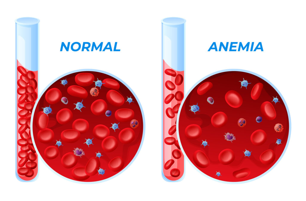
Autoimmune thrombocytopenia purpura: platelet count less than 20,000/mm3 Adequate or increased marrow megakaryocytes Presence of detectable platelet associated immunoglobulins not due to alloreactive antibodies or posttransfusion purpura Prior response to immunosuppressive therapy Platelet count chronically less than 20,000/mm3 with petechial bleeding or less than 50,000/mm3 with other bleeding OR Any history of life threatening hemorrhage Refractory to conventional therapy for at least 21 days Splenectomy At least 1 additional immunosuppressive therapy applied after splenectomy OR Controlled on conventional therapy but at price of unacceptable toxicity: Serious steroid related toxicity Absolute neutrophil count less than 500/mm3 25% of time, pure red blood cell transfusion dependent or other toxicities (e.g., hemorrhagic cystitis) that are a consequence of chronic or cytotoxic therapy Unable to wean from chronic daily or intermittent cytotoxic therapy Autoimmune hemolytic anemia or pure red cell aplasia, AIHA: Hemolytic anemia Hemoglobin less than 10.0 g/dL without transfusion Hemolysis as evidenced by both: Sustained reticulocytosis (greater than 125,000/mm3) without evidence of active bleeding or increasing hemoglobin Laboratory evidence of hemolysis Positive direct antiglobulin test or equivalent immune adherence test No evidence for paroxysmal nocturnal hemoglobinuria Negative Ham’s test and sucrose hemolysis. For PRCA: Anemia due to selective decrease in marrow erythroid precursors Hemoglobin less than 10.0 g/dL without transfusion Severe reticulocytopenia (less than 20,000/mm3 despite anemia) Severely decreased marrow erythroid precursors Positive marrow coculture with serum or cells or response to immunosuppression No evidence for PNH Negative Ham’s test and sucrose hemolysis Severe disease: Chronic (i.e., greater than 1 year) Transfusion dependent or untransfused hemoglobin less than 8.0 g/dL Ferritin greater than 2,000 or evidence of organ dysfunction due to iron overload Refractory to conventional therapy after all 3 of the following: High dose steroids (at least 1 mg/kg) for at least 21 days Splenectomy (except cold reactive antibodies) 1 additional immunosuppressive therapy OR Controlled on conventional therapy but at price of unacceptable toxicity Rheumatoid arthritis: Morning stiffness for at least 6 weeks Arthritis of 3 or more joint areas Arthritis of hand joints Symmetric arthritis Rheumatoid nodules Serum rheumatoid factor Radiographic changes Active rheumatoid disease as evidenced by all of the following: Elevated Westergren erythrocyte sedimentation rate Minimum of 16 swollen or tender joints using the 28 joint count method Must be at high risk for developing deforming joint disease as defined by at least 2 of the following: High titer IgM-IgG rheumatoid factor Radiographic evidence of erosive arthritis developing within the first 24 months of clinical disease Functional class II or III Refractory to conventional therapy after 12 months of: Methotrexate used in combination with cyclosporine, hydroxychloroquine, or sulfasalazine OR Intramuscular gold therapy (total dose greater than 1.0 g and duration at least 6 months) OR Controlled on conventional therapy but at price of unacceptable toxicity Juvenile rheumatoid arthritis: Under 16 years of age at onset Arthritis in 1 or more joints as defined by swelling or effusion, or presence of 2 or more of the following: Limitation of range of motion Tenderness or pain on motion Increased heat Duration of disease 6 weeks or longer Onset type defined by type of disease in first 6 months: Polyarthritis (i.e., 5 or more inflamed joints) Oligoarthritis (i.e., less than 5 inflamed joints) Systemic (i.e., arthritis with characteristic fever) Exclusion of other forms of juvenile arthritis Active disease evidenced by 1 of the following: Minimum of 2 swollen or tender joints using the 71 joint count method Endocardial or myocardial disease, or serositis Anemia or thrombocytosis of chronic disease High risk for developing deforming joint disease or evidence of potential life threatening involvement for at least 1 internal organ system Radiographic evidence of erosive arthritis developing within first 24 months of clinical disease Functional class II or III Endocardial, myocardial, pericardial, and/or pleural disease Hemoglobin less than 10.0 g/dL or platelet count greater than 600,000/mm3 Refractory to conventional therapy after 12 months of methotrexate used in combination with hydroxychloroquine, sulfasalazine, azathioprine, cyclosporine, or cyclophosphamide OR Controlled on conventional therapy but at price of unacceptable toxicity Systemic lupus erythematosus: Malar rash Discoid rash Photosensitivity Oral ulcers Arthritis Serositis Renal disorder Neurologic disorder Hematologic disorder Immunologic disorder Antinuclear antibody Must have at least 4 of 7 variables on the lupus activity scale measured Evidence of potential life threatening involvement of at least 1 internal organ system Endocardial and/or myocardial disease Central nervous system disease Pulmonary parenchymal disease Renal disease defined as WHO III, IV or V and a high activity and low chronicity index Immune mediated cytopenias Refractory to conventional therapy after attempts to control disease with at least 2 drugs, including prednisone and 1 of the following: Azathioprine Cyclophosphamide (greater than 500 mg/m2 monthly for 6 months) Cyclosporine OR Controlled on conventional therapy but at price of unacceptable toxicity Vasculitis Definitive diagnosis of 1 of the following forms: Churg-Strauss syndrome Giant cell arteritis Henoch-Schonlein purpura Hypersensitivity vasculitis Polyarteritis nodosa Takayasu arteritis Granulomatosis with polyangiitis (formerly known as Wegener’s granulomatosis) Evidence of active disease defined as reversible manifestations of the underlying inflammatory process Must have 1 or more of the following: Elevated Westergren erythrocyte sedimentation rate Elevated C reactive protein Decrease serum complement levels Evidence of potential life threatening involvement of at least 1 internal organ system Endocardial and/or myocardial disease Central nervous system disease Pulmonary parenchymal disease Renal disease defined as WHO III, IV or V and a high activity and low chronicity index Immune mediated cytopenias Refractory to conventional therapy (i.e., failed or relapsed within 6 months) after attempts to control disease with at least 2 drugs, including prednisone and 1 of the following: Methotrexate Azathioprine Cyclophosphamide Cyclosporine OR Controlled on conventional therapy but at price of unacceptable toxicity Performance status: ECOG 0-1 ECOG 2 allowed provided symptoms directly related to autoimmune disease Hepatic: No history of severe, prior or ongoing chronic liver disease Bilirubin less than 2.0 mg/dL AST less than 2 times upper limit of normal (ULN) Alkaline phosphatase less than 2 times ULN Renal: Creatinine less than 2.5 mg/dL OR Creatinine no greater than 2 times normal baseline for age in pediatric patients Cardiovascular: No symptoms of cardiac disease No active ischemic heart disease Ejection fraction greater than 45% by MUGA No uncontrolled hypertension Pulmonary: FEV1/FVC at least 60% OR Resting PO2 at least 80 mm Hg DLCO greater than 50% predicted O2 saturations greater than 94% in children unable to perform PFTs Neurologic: No active or ongoing ischemic or degenerative CNS disease not attributable to underlying disease Other: Not pregnant No poorly controlled diabetes HIV negative
Total Enrolment
10
Contact Details
Fairview University Medical Center, Minneapolis, Minnesota, 55455, United States
All content and media on the HealthEngine Blog is created and published online for informational purposes only. It is not intended to be a substitute for professional medical advice and should not be relied on as health or personal advice. Always seek the guidance of your doctor or other qualified health professional with any questions you may have regarding your health or a medical condition. Never disregard the advice of a medical professional, or delay in seeking it because of something you have read on this Website. If you think you may have a medical emergency, call your doctor, go to the nearest hospital emergency department, or call the emergency services immediately.
Related Articles


Need a health appointment?
Find and book a doctor, dentist, physio and more on Healthengine
Find a practitioner