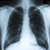
Do preconceived expectations impact on doctor analysis of x-rays?
Scientists have long suspected that clinicians’ ability to read x-rays can be skewed according to what they expect to find, but a University of Sydney study published in the international journal Radiology did not find evidence to support this theory.
Lead author Warren Reed from the Faculty of Health Sciences put 22 highly experienced radiologists from the American Board of Radiologists to the test by studying the impact of prevalence expectation on the ability of the radiologists to detect pulmonary nodular lesions, or lung cancer.
The 22 radiologists were divided into three groups and given an identical set of 30 chest x-rays to examine. Researchers tested whether accuracy rates improved or declined if the groups of radiologists were given different information about the number of cancer nodules present in the x-rays, thus altering their expectations about what to find.
The findings of the study show that there was no significant change in the number of cancer nodules that observers noticed after being given a prior expectation. However the study found significant increases in eye movements that demonstrate that efficiency decreases at higher prevalence expectations rates.
“What tended to change was the way they looked at the images,” Dr Reed said.
“This is an important finding as it could lead to extra fatigue which is detrimental to performance of radiologists, especially during long reporting sessions,” Dr Reed said. “It was quite a revelation. You’d assume that a radiologist would sit down and look at things in the same way, time and time again.”
The study is of particular importance to those interested in studying the relationship between observer process and observer accuracy under laboratory and clinical conditions.
Dr Reed is a member of the Image Optimisation and Perception Group in the Faculty of Health Sciences, which researches image perception and interpretation of medical imaging. Biological imaging is a fundamental component of health care systems and laboratory research. In the wake of major technological advancements the area is undergoing unprecedented change whereby imaging techniques, along with analytical tools, are being developed to display biological tissues and processes in new ways.
(Source: University of Sydney: Radiology)
More information
 | For more information about x-rays, including how they work, preparing for an x-ray, what happens during an x-ray and risks and benefits of x-rays, see X-Ray (General Radiology and Fluoroscopy). |
Dates
Tags
Created by:

 Login
Login


