Staging of Skin (Cutaneous) Melanoma
Cancer staging
Staging is a term used to define a simplified method of describing the extent of a cancer. Staging of cancers is important as it:
- Helps guide appropriate treatment;
- Can be used to guide prognosis;
- Can identify clinical trials that may be a suitable treatment option for a patient; and
- Allows a common terminology for exchange of information as well as evaluation and comparison of clinical trials.
The staging of your cancer is performed by your treating health professional, which may include an oncologist (doctor who specialises in cancer) if you need to be referred to one.
Staging is performed at the time, or soon after, the diagnosis has been made. Therefore the stage of your cancer reflects the extent of your cancer at the time of diagnosis. It does not change overtime. If your cancer changes over time these changes can be added on to, but never replace, the stage that you were given at diagnosis. In very rare situations your cancer may be restaged (i.e. you have all of the tests to stage your cancer again) and when this is done a little ‘r’ is put in front of the stage to reflect that it has been restaged and is not the stage given at diagnosis.
What is the TNM staging system?
The most widely used staging system for skin (cutaneous) melanoma is the TNM system. This system groups cancers into stages based on:
- Tumour (T) – the size and/or extent of the primary tumour;
- Nodes (N) – if there is any spread to nearby lymph nodes (collections of cells that act as a filter for the immune system); and
- Metastases (M) – the presence of metastasis (spread of the tumour through the bloodstream to distant sites, for example melanoma cancer cells spreading through the blood to create a new tumour in the lungs or other site).
Cancer staging systems are revised regularly to reflect the current medical knowledge available. The most recent revision of the cutaneous (skin) melanoma staging system is the 7th edition of the Cancer Staging Manual, and applies to all cancers diagnosed on or after 1 January 2010.
The TNM staging categories for cutaneous melanoma are shown in Table 1 below.
Table 1. TNM staging categories for cutaneous melanoma.
| Classification T | Thickness (mm) | Ulceration status/Mitotic rate |
| Tis | NA | NA |
| T1 | <1.00 | a: without ulceration* and mitosis** <1/mm2 b: with ulceration or mitoses >1/mm2 |
| T2 | 1.01-2.00 | a: without ulceration b: with ulceration |
| T3 | 2.01-4.00 | a: without ulceration b: with ulceration |
| T4 | >4.00 | a: without ulceration b: with ulceration |
| N | No. of Nodes with cancer present | Burden of cancer spread to the nodes |
| N0 | 0 | NA |
| N1 | 1 | a: micrometastasis^ b: macrometastasis# |
| N2 | 2-3 | a: micrometastasis b: macrometastasis c: intransit metastases+ / satellites without metastatic nodes |
| N3 | 4+ or matted nodes or intransit metastases+ / satellites with metastatic nodes | |
| M | Site | Serum LDH = |
| M0 | No distant metastases | NA |
| M1a | Distant metastases to the skin, subcutaneous tissue or lymph nodes | Normal |
| M1b | metastases to the lung | Normal |
| M1c | metastases to all other internal organs Any distant metastasis | Normal Elevated |
| *ulceration refers to an area of skin that has been eroded away and may bleed, itch or form a scab **mitosis is the process in which melanoma cells divide and replicated to form more cells ^ micrometastasis is where evidence of cancerous cells in the lymph nodes is made only after biopsy # macrometastasis is where there is evidence of cancerous cells in the lymph nodes clinically (i.e. increase in size, hard to touch, generally painless) that is then confirmed by biopsy + intransit metastases / satellites refers to cancers that have spread and begin to grow more than 2 centimeters away from the primary tumor but have not yet reached the nearest lymph node/s = Serum LDH is a substance that is measured in the blood and has been shown to be important in the prognosis of melanoma that has spread to distant sites | ||
Tumour
The first section of the TNM staging system refers to the primary tumour. The classification as Tis, T1, T2, T3 or T4 (in the left hand column of Table 1) depends upon the thickness of the melanoma, if there is any ulceration to the melanoma site and the rate of mitosis (this is a scientific term to describe the rate at which the cells divide to grow more cells / replicate). In melanoma patients it has been shown that as the rate of mitosis increases, the rate of survival decreases.
As an example, a melanoma that is 1.5mm thick that has some ulceration (where an area of the skin has been eroded away and may bleed or itch or form a scab) would be classified as T2b.
Nodes
The second component of the TNM staging system refers to the involvement of lymph nodes (collections of cells that act as a filter for the immune system). The classification increases as the number of lymph nodes involved increases. For example, if there is only one lymph node affected, the classification would be N1 compared to if 2-3 lymph nodes were affected then the designation would be N2. These can be further classified depending on whether the spread is micro- (there are too few cancer cells to be picked up by screening or diagnostic tests) or macro-metastasis.
Metastases
The final component of the TNM staging system is dependent on whether there is any evidence for spread to other places in the body such as other skin sites, the lung or other organs within the body.
Final grouping
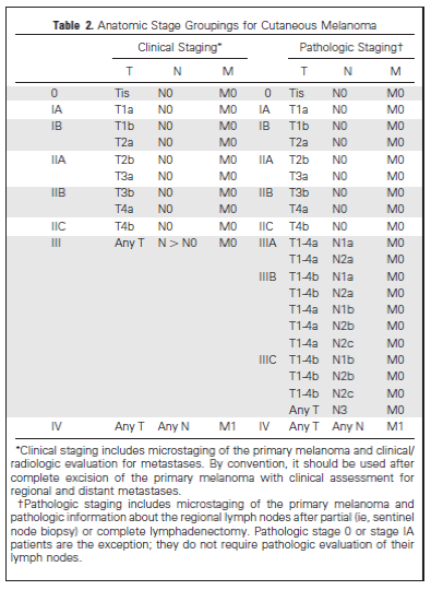
Once information is gathered pertaining to T, N, and M, this can then be used to determine the stage of the cutaneous melanoma as outlined below:
Table 2. Anatomic stage groupings for cutaneous melanoma.

As an example, stage 1A melanomas are those that:
Are <1mm thick, have no ulceration and a mitotic rate less than 1/mm2 (T1a) and has not spread to lymph nodes (N0) or other distant sites (M0).
In comparison, a stage IV melanoma is one that can be any thickness, have ulceration or not, have any mitotic rate, have spread to any number of lymph nodes and has metastasised (spread) to distant sites. Note that for this stage the important part of the staging is the fact that it has spread to distant sites.
A melanoma that has spread to the lymph nodes, but not other distant sites, is stage III. There are a number of T and N classifications within this stage that are grouped into either IIIA, IIIB or IIIC.
Kindly written and reviewed by Dr Allison Johns Bsc (Hons) MBBS, Doctor at Child and Adolescent Health Services, Women and Newborns Health Service and Editorial Advisory Board Member of Virtual Medical Centre.
References
- National Cancer Institute. Cancer Staging (online). March 2013 [Accessed 24/10/14]. Available from [URL Link]
- Balch CM, Gershenwald E, Soong S, Thompson JF, Atkins MB, Byrd DR, Buzaid AC, Cochran AJ, Coit DG, Ding S, Eggermont AM, Flaherty KT, Gimotty PA, Kirkwood JM, McMasters KM, Mihm MC, Morton DL, Ross MI, Sober AJ and Sondak VK. Final version of 2009 AJCC Melanoma Staging and Classification. J Clin Oncol 2009; 27: 6199-6206. [Abstract] [Full text]
- Cancer Council Australia /Australian Cancer Network / Ministry of Health, New Zealand. Clinical Practice Guidelines for the Management of Melanoma in Australia and New Zealand (online). October 2008 [Accessed 24/10/14]. Available from [URL Link]
- American Society of Clinical Oncology. Stages of Cancer (online). 2014 [Accessed 12/11/14]. Available from [URL Link]
- National Cancer Institute. NCI Dictionary of Cancer Terms: in-transit metastasis (online). No date [Accessed 16/11/2014]. Available from [URL Link]
Dates
Tags
Created by:

 Login
Login


