 Login
LoginRadionuclide (Isotope) Scan

- Introduction to radionuclide scans
- Why are radionuclide scans performed?
- Preparing for a radionuclide scan
- What happens during a radionuclide scan?
- Risks and complications of radionuclide scans
Introduction to radionuclide scans
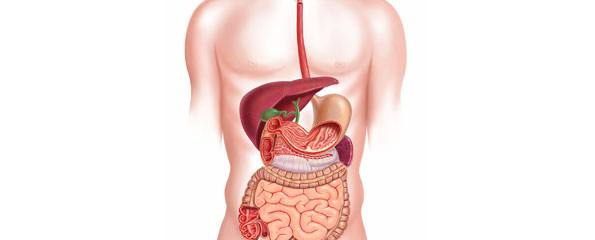
A radionuclide scan (also known as a radioisotope scan) is an imaging technique used to visualise parts of the body by injecting a small dose of a radioactive chemical into the body.
These chemicals localise to specific organs and tissues depending on the type of substance used and then emit small beams of radiation (called gamma rays) that can be detected by the gamma camera. Radionuclide scans are used in various fields of medicine such as identifying areas of infection or excess bone turnover. Bone scans and thyroid scans are common examples of radionuclide scans. In the gastrointestinal tract they can be used to identify sites of bleeding, measure the extent of inflammation and assess the movement of food substances through the stomach.
Why are radionuclide scans performed?
Radionuclide scans show the size, shape, position, and some function of the target organs that take up the particular chmeical injected into your body. Different chemicals are often used to visualise different organs and tissues.
Your doctor may suggest this investigation for a number of problems:
- Gastrointestinal bleeding: If you are noted to have recurrent bleeds from the gastrointestinal tract, this scan can help to find the source of bleeding when other techniques such as endoscopy have failed. To do this your red blood cells are labelled with a substance called technetium and areas of bleeding will show up as ‘hot spots’ on the monitor.
- Inflammatory bowel disease: The extent of inflammation in Crohn’s disease or Ulcerative colitis can be measured by this scan. This may help your doctor decide which treatment is most suitable for your condition.
- Gastric function: when certain labels are applied to bowel contents the rate of gastric emptying and any degree of reflux can be determined.
- Hepatobiliary disease: A particular chemical is used which is taken up by liver cells and excreted into bile. Recall bile is a thick, yellow substance containing bile salts which help absorb fats from the intestines. The pattern of uptake can identify if ceratin bile ducts are obstructed which may required treatment.
You should recognise that whilst this investigation can show some of these problems, it doesn’t provide very accurate detail of the structures it examines. Sometimes further investigations may be required in order to make a diagnosis.
Preparing for a radionuclide scan
- The person examining you may take a detailed history about your health and medications. It is important they know all the drugs you are taking and which previous contrast investigations (e.g. barium enema) you have had as certain substances can interfere with the isotopes given.
- They will explain the procedure in detail including the reason you are having this investigation and the risks. You will need to sign an informed consent form.
- Depending on the chemical used you may be required to fast overnight before the investigation.
- You must remove all jewellery before you are scanned as metal can block the radiation rays.
What happens during a radionuclide scan?
Prior to the investigation a small amount of the appropriate radionuclide chemical will be injected into the vein of your arm. You will feel a slight prick when the needle is inserted. This is then given time to spread to the organ or tissue that needs to be examined (e.g. your bowel, liver or spleen). Once in the tissue, active cells will take up the chemical and emit gamma rays that can be detected by the gamma camera. This forms an image on a television screen which can be interpreted by the examiner.
During the scan you will be required to remain very still lying down so the gamma cameras can detect the radiation emitted and get accurate images. The time taken for the investigation varies according to the site examined and the substance used. You may be encourage to drink plenty of water following the scan to allow the kidneys to remove the chemicals form your body.
Risks and complications of radionuclide scans
Overall radionuclide scans are considered a fairly safe procedure. They are non-invasive and cause little patient discomfort. However, like x-ray and other investigations they expose the body to a small dose of radiation. This can damage cells and cause mutations particularly in growing fetuses. Therefore if you are pregnant you must be made aware of the potential risks to your baby. In addition there is also a small risk that you may be allergic to the chemicals used for the scan. In patients sensitive to the chemicals nausea, vomiting and headaches can occur.
References
- Burkitt, Quick. Essential Surgery. 3rd Edition.Churchill Livingstone. 2002.
- Khilnani N, Hussain N, Gastrointestinal Bleeding, Emergency Medicine 2005: 37(10); 27-32.
- Kumar, Clark. Clinical Medicine. 5th Edition. Saunders. 2002.
- Medline Plus- Medical Encyclopedia, Isotope Study, 2005. Available [online] at URL: http://www.nlm.nih.gov/medlineplus/ency/article/003827.htm
- Mitchell S, Schaefer D, Dubagunta S, A New View of Occult and Obscure Gastrointestinal Bleeding, American Family Physician 2004;69:875-81.
- PatientUK- Radionuclide (Isotope) Scan, EMIS and PIP, 2003. Available [online] at URL: http://www.patient.co.uk/showdoc/27000283/
Dates
Tags
Created by:


