Genetic typing of cancers
- Introduction
- Biological pathways in genetic cancers
- Biologically targeted therapies
- Conclusion
- References
Introduction
Individuals that inherit a genetic mutation from their parents are at a greater risk of certain types of cancers. Familial cancer syndromes are the group of cancers where patients inherit genes that do not function properly, increasing the risk of developing cancer.
Approximately 100 familial cancer syndromes have been identified and this number is growing. The genes identified as increasing the risk of cancer are usually autosomal (mutations that occur on non-sex chromosomes) dominant (only one of the pair of chromosomes has to have the mutation in order for it to be expressed) although they can also be autosomal recessive (both chromosomes must have the mutation for it to be expressed). The identification of these genes has improved diagnosis and helped identify new cancer treatments.
This article explores which genes are involved in the most common familial cancer syndromes, the genetic markers of disease severity and prognosis (how a disease will develop over time), and the role of genetic typing of cancers in selecting an appropriate treatment, such as biologically targeted therapies.
Biological pathways in genetic cancers
 Normal cell growth and function is controlled by growth factors (natural agents that stimulate cell growth) that bind to receptors on the cell membrane. These receptors set off a cascade of molecules (in a process controlled by enzymescalled tyrosine kinases) to transmit the signal to the cell nucleus. Once in the nucleus, the signal initiates the production of certain proteins based on the genetic code in the cell’s DNA. These proteins have a wide range of functions, including cell apoptosis (cell death). If a cell is malfunctioning, it can be induced to self-destruct by stimulating these apoptosis pathways. This is an important element of the body’s defence against cancer, by which cells that have cancer-causing potential can be quickly eliminated. Familial cancer syndromes emerge when mutations in genes (or in associated proteins) cause this self-regulating process to be disrupted, potentially leading to the uncontrolled cell growth known as cancer.
The two types of genes relevant to familial cancers are known as oncogenes and tumour-suppressor genes. Oncogenes are genes that regulate cell growth and cell differentiation (the process by which stem cells become specific types of cells such as skin cells, muscle cells etc.); acquiring genetic mutations can alter their function and activity in such a way as to promote the development of cancer. These changes result in abnormal structure and function in transcribed proteins essential to cell growth and differentiation. Tumour-suppressor genes normally regulate cell division and initiate apoptosis if there is damage to DNA. Genetic changes in tumour-suppression genes lead to a loss of this regulatory role and can lead to uninhibited cell growth, such as in cancer.
Inheriting a genetic mutation in a tumour-suppressor gene does not automatically cause cancer; rather, it increases the likelihood of certain types of cancers emerging. Individuals without inherited genetic mutations are still able to develop these cancers; however, they are significantly less likely to occur. Most of the familial cancer syndromes are autosomal dominant mutations of tumour suppressor genes.
The study of genetic mutations in familial cancers has also helped discover treatment targets for individuals who develop the same cancer without having genetic mutations. Genetic testing can determine if patients have a hereditary or non-hereditary (sporadic) form of a cancer and can therefore be given the appropriate treatment.
Breast and ovarian cancer
BRCA-1 and BRCA-2
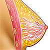
The BRCA (BReast CAncer) genes are a common example of a genetic mutation increasing the risk of cancer. An estimated 0.2%–0.33% of the population have mutations in the BRCA1 or BRCA2 genes, but this value can vary greatly between races. It is as high as 8.3% in some Ashkenazi Jewish women. However, BRCA mutations are estimated to account for 5–10% of breast cancers and 10–15% of ovarian cancers in Caucasian women.
BRCA1 or BRCA2 mutations in women are associated with a lifetime risk of breast cancer of up to 85%, and up to 26% for ovarian cancer. There is also evidence that BRCA mutations are associated with increased risk ofprostate cancer, pancreatic cancer, gallbladder and bile duct cancer, stomach cancer, malignant melanoma and fallopian tube carcinoma.
Guidelines recommend that women who have a positive family history of breast or ovarian cancer should be referred for genetic counselling and evaluation by BRCA testing.
Human epidermal growth factor receptor 2 (HER2)
Human epidermal growth factor receptor 2 (HER2) is a protein transcribed from the ERBB2 gene that stimulates growth in many cell types. However, mutations in the ERBB2 gene can cause over-expression of HER2.
Over-expression of HER2 occurs in 15–30% of breast cancer cases and is strongly associated with increased disease recurrence and worse prognosis (when non-specific chemotherapy isn’t used). It is also associated with an increased response to certain types of breast cancer therapies and a poorer response to other therapies.
Testing for HER2 is recommended for all women with primary invasive breast cancer. By identifying HER2-positive patients, breast cancer treatment plans can be adjusted to ensure the most effective therapy is given.
 | For more information about the risk factors, epidemiology and treatment of breast cancer, as well as videos and animations, see Breast Cancer. |
Renal cancer
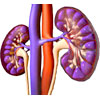
 Renal cell cancers (known as renal cell carcinomas, or RCCs) are derived from certain cell types in the renal cortex. RCC accounts for 2–3% of all cancer in adults worldwide. RCCs are very often associated with predisposing hereditary syndromes or genetic mutations, although risk factors such as BMI, high blood pressureand cigarette smoking predispose to its development.
The hereditary diseases that have a high risk of RCCs (called the renal cell cancer syndromes) include:15
- von Hippel-Lindau (VHL) syndrome: Patients with VHL syndrome have a rare autosomal dominant mutation in the VHL gene, which is a tumour-suppressing gene. This increases the risk of developing hemangioblastomas (cancers of the central nervous system) in the cerebellum, spinal cord, kidney andretina.
- C-met proto-oncogene mutation in hereditary papillary renal carcinoma (cancer in a specific part of the kidney).
- Hereditary leiomyomatosis renal cell carcinoma (HLRCC) is a hereditary cancer syndrome associated with the development of skin and uterine leiomyomas (abnormal mass in muscle tissue) and aggressive papillary renal carcinomas.
- Birt-Hogg-Dubé (BHD1) gene mutation: A hereditary cancer syndrome due to a loss of function of the BHD tumour suppressor gene. Affected individuals are at risk for the development of hair follicle fibrofolliculomas (small papules usually located on the head neck or upper body), pulmonary cysts (fluid-filled sac in the lung) and renal tumours.
- TSC1 or TSC2 gene mutations can cause tuberous sclerosis, a hereditary syndrome associated withcysts and malignant tumours of the heart, kidneys, pancreas and peritoneal cavity.
 | For more information about renal cancer including treatment, statistics and more, see Renal Cancer. |
Bowel cancer
Familial adenomatous polyposis (FAP)
FAP is an autosomal dominant syndrome of mutations in the APC tumour suppressor gene. This condition causes patients to develop hundreds to thousands of benign tumours (adenomas) in the colon, several of which will likely acquire further abnormalities and develop into a true bowel cancer. Approximately 50% of people with FAP develop adenomas by age 15 and 95% by age 35. FAP accounts for about 1% of annual colorectal cancer in the United States.
Hereditary non-polypoid colorectal cancer (HNPCC)
 HNPCC is an autosomal dominant cancer syndrome caused by mutations in DNA repair genes and accounts for 6–15% of colorectal cancers. It is believed that HNPCC tumours arise from a small number of adenomas that have a higher potential for becoming malignant due to these mutations. These mutations commonly occur in the genes MSH2, MLH1, MSH6 and PMS2. These mutations are also associated with an increased risk of endometrial, ovarian, pancreatic and gastric cancers.
By age 70, nearly 90% of HNPCC cases can be attributed to mutations in MSH2 and MLH1. Individuals with a newly diagnosed colorectal cancer should be screened for HNPCC genetic mutations.
Gastrointestinal stromal tumours (GIST)
Gastrointestinal stromal tumours (GIST) are the most common mesenchymal tumours of the gastrointestinal tract, with an estimated annual incidence of 1.1 to 1.5 cases per 100,000 people. Mutations in the KIT gene cause the enzyme tyrosine kinase to malfunction and over-activate, affecting intra-cellular signalling for cell growth and increasing the risk of GIST.
Understanding this molecular pathway has led to the development of the drug imatinib (sold as Glivec), a potent tyrosine kinase inhibitor, in the treatment of GIST.
 | For more information about the different types of bowel cancer and how they are detected and treated, see Bowel Cancer (Colorectal Cancer). |
 | For more information about the different types of stomach cancer including statistics, treatment and more, see Stomach Cancer. |
Prostate cancer
Hereditary prostate cancer (HPC)
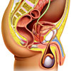
The genetics of hereditary prostate cancer are more complex than that of other cancer types. Research suggests that dominantly inherited susceptibility genes cause 5–10% of prostate cancers and up to 40% of early-onset prostate disease. While diagnosed earlier (approximately 7 years earlier on average), hereditary prostate cancer does not differ clinically from other, non-hereditary prostate cancers. Multiple genetic mutations have been studied (including the HPC1 gene) and multiple genetic variants have shown only modest increases in prostate cancer risk.
 | For more information about how prostate cancer is detected and treated, see Prostate Cancer. |
Multiple endocrine neoplasia 1 and 2
Multiple endocrine neoplasia (MEN-1 and MEN-2) syndromes are disorders that greatly increase the risk of developing multiple tumours in endocrine glands. Individuals with MEN-1 develop primary hyperparathyroidism, neuroendocrine tumours of the pancreas, stomach, duodenum, bronchus and thymus, pituitary tumours and adrenal adenomas. MEN syndromes are caused by mutations in MEN-1 tumour suppressor genes and are inherited in an autosomal dominant manner. The MEN-1 gene encodes a protein called menin that regulates gene activity and can lead to tumour development if it’s function is disturbed.
Retinoblastoma
Retinoblastoma is a malignant eye tumour of childhood (usually < 5 years of age) that can affect one or both eyes and can cause visual loss. Most cases of retinoblastoma are hereditary, and are associated with retinoblastoma gene (Rb gene) deletion. These mutations are autosomal dominant. Individuals with RB1mutations are also at risk of other tumours outside the eye.
MicroRNAs and cancer
MicroRNAs (miRNA’s) are small RNA sequences, like a small segment of a single strand of DNA. MiRNA’s are involved in gene regulation and over 300 miRNA genes have been identified in the human genome.
Various miRNAs have been found to be abnormally expressed in several human cancers and have been attributed to several mechanisms, such as chromosomal rearrangement, defects in the miRNA synthesis pathway, and regulation by other transcriptional factors. MiRNAs appear to contribute to cancer development due to their role in oncogene signalling pathways. There is evidence that the expression of miRNA can help distinguish a cancer cell’s lineage, confirm cancer diagnoses and predict outcomes for patients.
Studies have shown that different tumour cell lines share similar changes in miRNA expression, regardless of where the tumour originates. For example, the Let-7 family of miRNA is usually highly expressed in normal tissues while in multiple cancer cell lines, this expression is reduced.
Biologically targeted therapies
Understanding the molecular pathways of hereditary cancer syndromes has helped the development of biological therapies for cancer treatment, which are used more and more frequently for many cancer types. Whereas chemotherapies reduce cell replication (effective in inhibiting fast-dividing cancer cells), biological therapies target the molecular pathways in cellular growth specifically to reduce the growth and spread of cancers.
These therapies can be used alone or in combination with existing anti-cancer treatments. For example, the use of targeted biological therapies has revolutionised the treatment of renal cell carcinoma (RCC), through the use of multiple receptor tyrosine kinase (rtk) inhibitors such as sunitinib (Sutent) and sorafenib (Nexavar), and mammalian target of rapamycin (mTOR) inhibitors, such as sirolimus (Rapamune), temsirolimus (Torisel) and everolimus (e.g. Afinitor, Certican).
By determining the underlying genetic mutations in an individual’s cancer, it is possible to determine which biological therapy will be most effective and “personalise” treatment. This has been demonstrated in the use of the anaplastic lymphoma kinase (ALK) inhibitor crizotinib (Xalkori) in certain cancers. Mutations in ALK are seen in subsets of patients with non-small cell lung cancer (NSCLC), as well as anaplastic large-cell lymphoma and neuroblastoma. Crizotinib is used in these cancer types as it suppresses cell lines with ALK activation due to genetic mutations. Patients without this mutation do not benefit from crizotinib use. By determining the patient’s ALK status, crizotinib can be used or withheld to improve clinical outcomes for patients.
Conclusion
There is an increasing recognition of the role of genetic mutations and cellular signalling pathways in the development of hereditary cancer syndromes. Not only do these mutations offer insight into the development of both hereditary and non-hereditary forms of cancer, but they also provide potential therapeutic targets to prolong survival.
The growing use of biological therapies that affect cellular signalling pathways has revolutionised the treatment of cancer, a trend that will continue as treatment strategies can be personalised to individuals once genetic typing of their cancer is known.
Reference
- Garber JE. Hereditary Cancer Predisposition Syndromes. Journal of Clinical Oncology. 2005; 23: 276–292. [Abstract]
- Braunwald E, Fauci AS, Kasper DL, et al. Harrison’s Principles of Internal Medicine (15th edition). New York: McGraw-Hill Publishing; 2001. [Book]
- Knudson AG. Hereditary cancer: Two hits revisited. J Cancer Res Clin Oncol. 1996; 122: 135–140. [Abstract]
- Knudson A. Two genetic hits (more or less) to cancer. Nat Rev Cancer. 2001; 1 (2): 157–62. [Abstract]
- Nelson H, Huffman L, Fu R. Genetic risk assessment and BRCA mutation testing for breast and ovarian cancer susceptibility: systematic evidence review for the US Preventive Services Task Force Ann Intern Med. 2005; 143(5): 362–79. [Abstract | Full Text]
- John EM et al. Prevalence of Pathogenic BRCA1 Mutation Carriers in 5 US Racial/Ethnic Groups. JAMA.2007; 298: 2869–2876. [Abstract | Full Text]
- Campeau PM, Foulkes WD, Tischkowitz MD. Hereditary breast cancer: new genetic developments, new therapeutic avenues. Hum Genet. 2008; 124: 31–42. [Abstract]
- Cass I, Holschneide C, Datta N. BRCA-Mutation–Associated Fallopian Tube Carcinoma: A Distinct Clinical Phenotype? Obstet Gynecol. 2005; 106(6): 1327–34. [Abstract]
- Harris L, Fritsche H, Menne, R. American Society of Clinical Oncology 2007 update of recommendations for the use of tumour markers in breast cancer. J Clin Oncol. 2007; 25(33): 5287-312. [Abstract | Full Text]
- Tan M, Yu D. Molecular mechanisms of erbB2-mediated breast cancer chemoresistance. Adv Exp Med Biol. 2007; 608: 119-29. [Abstract]
- Wolff AC, Hammond ME, Schwartz JN et al. American Society of Clinical Oncology/College of American Pathologists guideline recommendations for human epidermal growth factor receptor 2 testing in breast cancer. Arch Pathol Lab Med. 2007; 131(1): 18-43. [Abstract | Full Text]
- Sturgeon CM, Duffy MJ, Stenman UH et al. National Academy of Clinical Biochemistry laboratory medicine practice guidelines for use of tumour markers in testicular, prostate, colorectal, breast, and ovarian cancers.Clin Chem. 2008; 54(12): e11-79. [Abstract | Full Text]
- Rini BI, Campbell SC, Escudier B. Renal cell carcinoma. Lancet. 2009; 373(9669): 1119-32. [Abstract]
- Chow WH, Gridley G, Fraumeni JF Jr. Obesity, hypertension, and the risk of kidney cancer in men. N Engl J Med. 2000; 343(18): 1305-11. [Abstract | Full Text]
- Pfaffenroth EC, Linehan WM. Genetic basis for kidney cancer: opportunity for disease-specific approaches to therapy. Expert Opin Biol Ther. 2008; 8(6): 779-90. [Abstract | Full Text]
- Curatolo P, Bombardieri R. Tuberous sclerosis. Handb Clin Neurol. 2008; 87: 129-51. [Abstract]
- Brosens LA, Keller JJ, Offerhaus GJ et al. Prevention and management of duodenal polyps in familial adenomatous polyposis. Gut. 2005; 54(7): 1034-43. [Abstract | Full Text]
- Cruz-Correa M, Giardiello FM. Familial adenomatous polyposis. Gastrointest Endosc. 2003; 58(6): 885-94. [Abstract]
- Bonadona V, Bonaïti B, Olschwang S et al. Cancer risks associated with germline mutations in MLH1, MSH2, and MSH6 genes in Lynch syndrome. JAMA. 2011; 305(22): 2304-10. [Abstract | Full Text]
- Evaluation of Genomic Applications in Practice and Prevention (EGAPP) Working Group. Recommendations from the EGAPP Working Group: genetic testing strategies in newly diagnosed individuals with colorectal cancer aimed at reducing morbidity and mortality from Lynch syndrome in relatives. Genet Med. 2009; 11(1): 35-41. [Abstract | Full Text]
- Blay JY, von Mehren M, Blackstein ME. Perspective on updated treatment guidelines for patients with gastrointestinal stromal tumours. Cancer. 2010; 116(22): 5126-37. [Abstract | Full Text]
- Bratt O. Hereditary prostate cancer: clinical aspects. J Urol. 2002; 168(3): 906-13. [Abstract]
- Liu H, Wang B, Han C. Meta-analysis of genome-wide and replication association studies on prostate cancer. Prostate. 2011; 71(2): 209-24. [Abstract]
- Pieterman CR, Vriens MR, Dreijerink KM et al. Care for patients with multiple endocrine neoplasia type 1: the current evidence base. Fam Cancer. 2011; 10(1): 157-71. [Abstract]
- Raue F, Frank-Raue K. Update multiple endocrine neoplasia type 2. Fam Cancer. 2010; 9(3): 449-57. [Abstract]
- Falchetti A, Marini F, Brandi ML. Multiple Endocrine Neoplasia Type 1. In: Pagon RA, Bird TD, Dolan CR, et al., editors. GeneReviews[online]. Seattle (WA): University of Washington, Seattle; 1993-2000 Aug 31 2005 [updated 2010 Mar 2]. [URL]
- Lohmann DR, Gallie BL. Retinoblastoma. In: Pagon RA, Bird TD, Dolan CR, Stephens K, editors. GeneReviews [online]. Seattle (WA): University of Washington, Seattle; 1993-2000 Jul 18 [updated 2010 Jun 10]. [URL]
- Hammond SM. RNAi, microRNAs, and human disease. Cancer Chemother Pharmacol. 2006; 58 Suppl 1: s63-8. [Abstract]
- Calin GA, Dumitru CD, Shimizu M et al. Frequent deletions and down-regulation of micro- RNA genes miR15 and miR16 at 13q14 in chronic lymphocytic leukaemia. Proc Natl Acad Sci. 2002; 99(24): 15524-9. [Abstract]
- Kapoor A. Inhibition of mTOR in kidney cancer. Curr Oncol. 2009; 16 Suppl 1: S33-9. [Abstract | Full Text]
- Yuan Y, Liao YM, Hsueh CT, Mirshahidi HR. Novel targeted therapeutics: inhibitors of MDM2, ALK and PARP. J Hematol Oncol. 2011; 4: 16. [Abstract | Full Text]
- Cimmino A, Calin GA, Fabbri M et al. miR-15 and miR-16 induce apoptosis by targeting BCL2. Proc Natl Acad Sci USA. 2005; 102(39): 13944-9. [Abstract | Full Text]
Dates
Created by:

 Login
Login


